| Structural Analysis of
Starvation Transmission Electron Microscopy is being
used to examine cells for structural alterations that are associated
with a transition to starvation. The TEM images can potentially complement
proteomics by providing visual clues into changes in protein-based
structures that might be reflected in the proteome, and broader changes
that reflect cell state, but may not be captured in the proteome analysis. |
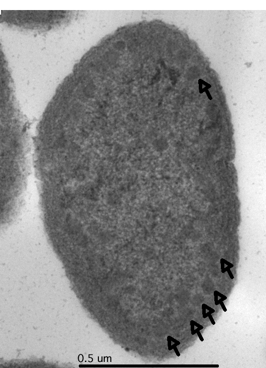
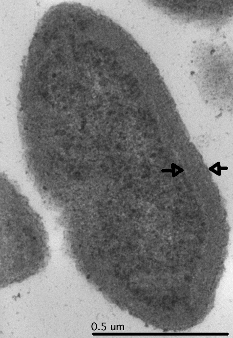
| Non-stressed, growing cells |
Larger
image
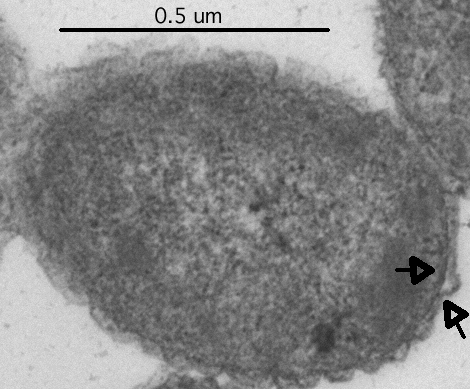
Possible carboxysome-like supramolecular aggregatesclustering near the
inner face of the cytoplasmic membrane
|
Larger
image
Multilamellar membranes, characteristic of N. europaea surround the
cells
|
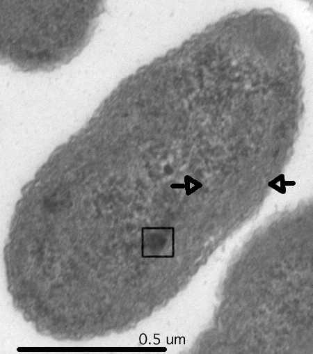
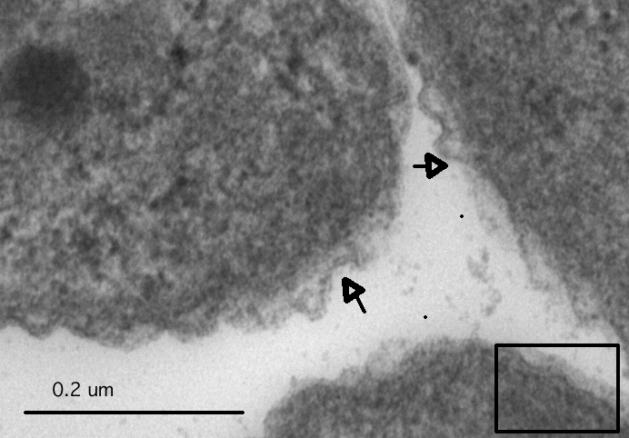
| Cells starved for ammonium for 2
weeks |
Larger image
Loosening of multilamellar membranes, electron dense bodies
form |
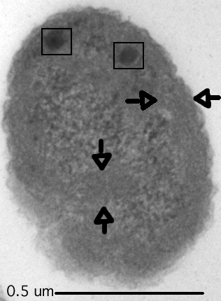
Larger image
Invagination of multilamellar membranes, multiple electron dense
bodies |
| Cells starved for ammonium
for 2 months |
Larger image
Multilamellar membranes degenerate |
Larger image
Zoom image of membrane degeneration TEM images produced in association with Randall Massey
(UW-Madison Medical College, Electron Microscopy Facility) |
Home
|